Showing posts with label neurology. Show all posts

02 April 2011

Overdue Update

I apologise for my absence. I was on rotation in downtown Detroit and the temporary crash-pad did not have internet (?!). I was on neurology, which is not at the top of my super-interesting-material list, but I did get to spend two weeks in the neuro-ICU and I do so love any kind of ICU. Bring me your super-sick, your actively dying and I will go to work with enthusiasm and diligence.


One of the wonderful things about this new hospital was the white chocolate macadamia cookies. And the Monday schwarma lunches. Yummy. Also interesting was editing my fellow's notes for proper English grammar and spelling (yes, I was called upon for spelling).

There was one very sad case: a patient who had a stroke at the young age of 41. We don't know why. He's now densely hemiplegic (can't move half of his body) and non-verbal. While under our care his wife found out she's pregnant. She's going to have a new baby and a husband in inpatient rehab who also needs her care. While rounding each morning he would start crying; he is cognitively intact and aware of his prognosis: he will likely not get much movement back.

In personal life news (yes, neurology is one of the rotations in which a life is possible), I've taken up P90X, which is quite challenging, but awesome. I finally had a good night out dancing, which I sorely needed. Sometimes there's just nothing like a cocktail and a good song to dance too. I even made some friends in Detroit, so hopefully I'll spend some more time exploring the city. Oh, and I signed up for motorcycle classes... shhh... don't tell my parents. It's going to be awesome when I visit them in May and hop on dad's Ducati! I'm working on signing up for tennis clinic, but it might be full. Boo.

Tomorrow morning is the beginning of Family Medicine... in Toledo. Also, the ramp up for studying for Step 2 (the second board exam, scheduled for June 2nd). Nevertheless, weekends off for another month so hopefully I'll be able to continue this whole "balance" thing.

09 March 2011

I feel dirty

I am on rotation at another hospital at the moment and one of our lecturers failed to show. Instead, I got to bond with some of my fellow students (for whom this hospital is home). One of them had a great horror story...


She was evaluating a patient in the ER who came in with priapism (a prolonged erection). Draining the organ was attempted, with no success. The student's attending then told her to "milk it." After some hesitation, she did as she was told. Another attending came by and asked her what on earth she was doing?! Turns out the first attending had been joking. The patient went on to surgery.

30 November 2009

Sounds like...

One of the blogs I read recently had a patient come in with Flea-bitis. It reminded me of a mother who brought her son in to neurology clinic for a second opinion. Apparently he had been diagnosed with something that sounded like Gardenias. The neurologist and the NP threw out a series of potential neurologic conditions... Guillian-Barre, maybe? After a quiet moment, I volunteered, do you mean Myasthenia Gravis? Yes, she did.

11 November 2009

Pearls of wisdom

Recent quotes from lecture:

"Our country handles schizoid personalities very well. That's why we have software engineers, pathologists and Montana." -Dr. J

"Biopsying an aneurysm is not a successful procedure." -Dr. G

"If someone's head is cut completely off, that's not an emergency - that's a tragedy." - Dr. G

03 November 2009

10 cent words

Medicine is full of big words with very specific meanings. Here is a fun paragraph from today:

Internuclear ophthalmoplegia (INO) is indicative of a particular opthalmoparesis. It is a disorder of conjugate lateral gaze in which the affected eye shows impairment of adduction. When the partner eye is abducted, it diverges from the affected eye. This produces horizontal diplopia. During extreme abduction, compensatory nystagmus can be seen in the partner eye. Convergence is generally preserved.

06 October 2009

Breaking bad news

A recent assignment for school caused me to recollect a patient from my previous hospital. An excerpt (the original essay is several paragraphs longer) from my assignment summarizes the story...

The patient, an 8-year-old boy, was the elder of two sons of a recently immigrated family. He had been admitted for status epilepticus, which proved to be refractory. He was ultimately placed in a medically induced coma (which he continued to seize through for a month) and the parents were asked whom else they would like present for a discussion of his prognosis.

The family requested that a doctor from their home country be involved and so a teleconference was established. Various members of the care staff reported their opinions and summarized the boy’s course to date. There was almost as much silence as there was talking and each person was careful to solicit and answer questions. The questions themselves guided the discussion. The parents really needed to believe that every option had been exhausted. They believed that because we had been able to find an etiology, we should be able to find a cure.

From a physician’s perspective, after a month of seizing and coma there was likely to be little brain function left. A multitude of testing revealed a genetic defect in a sodium channel, which was blamed for the seizure activity. The boy was not a surgical candidate because the seizures were multi-focal and originated from both hemispheres. Everything had been tried and nothing would break the seizures, which were still occurring roughly every three minutes. There was nothing more that could be done.

Most of the conversation centered on the futility of our treatments. That we, as doctors, could not even promise he would wake up if we took away the sedatives. Eventually the parents chose to withdraw support. I think they knew their decision from the moment they sat down; they just couldn’t say it out loud. I didn’t get the sense we had persuaded them; more that we gave them a safe and justified way of letting go and not feeling as if they were bad parents, that they were simply giving up because it was hard.

It was then, when they stated their decision, that I was so thankful for the private room (not a patient room) the whole conversation took place in. The family had somewhere they could be where they wouldn’t be disturbed or overheard. Not by a nurse who needed to take vitals, not by a doctor checking on another patient. They couldn’t hear the business of medicine still working around them, healing some of the other children who would eventually be able to go home to their parents. But the room also allowed us, as the medical staff, to separate the conversation we just had from the rest of our work. When you walk out the door and back onto the ward, you leave the heaviness in that room and focus on health and healing on the floor. The spatial separation aids the mental and emotional one.

That separation is not learned with one conversation. Or maybe it’s always imperfect. But I do know that I was unusually quiet the rest of the day. When a friend needed sympathy later that night, I just couldn’t muster any. His problems seemed so petty. The boy was only eight and he was dead. Dead because of a sodium channel, which seems like such a insufficient and small reason. He had a little brother who clearly didn’t understand what had happened. He had a mom and dad that somehow had to keep going. And while I wasn’t consciously dwelling on it, something in me didn’t let it go immediately. Within the week though, the petty problems regained their gravity and the pressing concerns of work and medical school applications took over.

- * - * - * - * - * -

I have to admit that delivering bad news became easier. Maybe not in the moment, but my recovery got much faster.

With regard to the family in the story, the little brother had ongoing issues coping with his brother's death. I personally think some of this had to do with the fact that the parents never allowed the younger son to visit the older one (they didn't want him to see his brother sick or in pain). Then again, I am neither a parent nor a psychologist, so I am not at all qualified on the subject.

19 June 2008

Reverse clinical pathology conference

Today I went to my first reverse clinical pathology conference. It's reverse because rather than starting with the patient's presentation and moving forward in time, you start with the pathology slides and try to reconstruct the symptoms and diagnosis based on what you see under the microscope.

There are pathology rooms with multi-headed microscopes: 8-10 viewing stations connected to a single slide mount so everyone is viewing the same slide simultaneously. A pathologist usually "drives" and physician and fellows (and me!) sit around the other viewers. The pathologist loaded in stained, prepared slides of the brainstem, cerebellum, and cortex for examination.

Slide #1 cerebellum. The cerebellar cortex is made up of three layers: the molecular layer, granule layer, and Purkinje layer. In our slide there was no surface granule layer, indicating the cells had already completed their migration and the patient must have been at least 12 months of age. The Purkinje cells were dying, a classic hypoxia sign. We also saw evidence of infection in the meninges (membranes covering the brain) and the parenchyma (brain tissue) such as macrophages and perivascular cuffing.

In the parenchyma we noted neuronophagia: dying neurons surrounded by microglial cells. This pointed us towards considering a viral diagnosis. Bacteria can stay in the meninges surrounding the brain, but viruses are obligate cellular parasites and thus more likely to appear in the neural tissue itself. We examined the neurons for inclusion bodies, which are accumlations (not exclusive to viral infections) within either the cytoplasm (ex rabies) or nucleus (ex. herpes). Not all viral infections have inclusion bodies, but their presence, absence and type can help to identify the specific pathogen. In this patient there were no inclusion bodies.

Slide #2: pons (brainstem). We noted extensive liquefactive necrosis, leaving the tissue with a spongelike appearance. Basically, the neurons are dying and pockets of fluid-filled space are left where there used to be brain tissue (necrosis = abnormal cell death). The extent of the damage in this patient was severe, indicating a rampant infection and acute, considering the cell types present.

Slide #3: midbrain (brainstem). The substantia nigra in the midbrain was noted to be unpigmented. This area usually develops pigment between 3-5 years of age (and can be lost again in old age as part of a neurodegenerative process). This placed the patient's age between 12 months and 3 years.

Slide #4: spinal cord. Most infections have a regional preference within the nervous system; they don't invade all areas equally. Thus, looking at which regions are affected can narrow down the pathogens under consideration. In our patient, the anterior horn of the spinal cord was disproportionately infected, which led us to consider the enteroviruses (ex polio, Hep A).

Conclusion: The patient turned out to be 25 months old and died of a massive Coxsackie viral infection. Coxsackie virus (type B pictured to the right) is probably best described as a non-paralytic polio and it can affect other organ systems besides the central nervous system, most notably the heart and eyes.

Note: this case has been previously published so the details have not been altered. All photos used are from searching google images and are not the slides from the actual conference.

20 April 2008

Reading medical memoirs

It is probably not surprising that throughout my post-bacc career I have been reading a series of medical memoirs, novels, and essays. There was House of God, which everyone reads at some point (and I have been told to re-read in residency), Better and Complications, both by Atul Gwande (he's kinda famous among the medical set), The End of Medicine (by a finance guy) and the Man who Mistook his Wife for a Hat (pop culture famous book on neurology by Oliver Sacks).

I recently started one by a female neurosurgeon, Katrina Firlik, and I am struck by how familiar it all is. I work in neurology, not neurosurgery, but I have seen many of the conditions she talks about. She didn't have to explain holoprosencephaly or hydrancephaly; I've seen them. I certainly don't have the knowledge base of peds neuro resident or even probably a well-educated, interested medical student, but I am conversant with the best of them on a limited subset of conditions.

The other theme that strikes me is that I have already begun the personal transformation that comes with being a physician. Dr. Firlik spends time explaining the sense of humor in the OR, the detachment of the physicians, the cold practicality that contributes to efficient care in times of crisis, but these paragraphs already ring hollow. They are exactly how I would explain it to someone on the outside, but there is really no way to make it ring true unless you've been there. Patients will never quite understand how you can tell them the worst news of their life and then spend an enjoyable afternoon hiking.

There are lot of things you don't realise when you start down this road to become a doctor, but this one might be the biggest, the most subtle, and the most significant. There is no undoing the change in how you view people and sickness; in this one way you will forever be apart from your non-medical peers.

18 April 2008

Leashes on kids

I must admit that subscribe to the idea best articulated on the Simpsons, "the leash demeans us both." But last Friday I met a mom who admitted to using a leash for her toddler and if I were her I would probably use one too.

Her child has a neurologic condition that, among other things, leaves her son both developmentally delayed and non-verbal. What this means is that while her son can walk and run and jump, he does not speak and has not yet learned common social cues.

The mom is clearly an involved and attentive mother, but as any mom or even babysitter can attest it is impossible to be watching every second of every day. What happens if she's paying for the groceries or using an ATM and he runs off? He can't say his name or who his mom is. He can't say where he lives or where he last saw his mom and we're not certain he would understand to look where he last saw her or follow a command given over a PA system.

Mom has sewn his name and address into all his clothes, just in case. She can't give him an ID necklace because he could hurt himself with it. He had a bracelet but he broke it - and the one before that. She's saving up to get one in a metal he can't break. What happens when he becomes coordinated enough to undo the clasp on it himself?

I don't think leashes are appropriate for most children, but I understand the trade-off here. I would also rather be the mom who gets stared at than the mom who loses her child.

10 April 2008

Wiki project done!

I recently finished one of the cooler projects I've ever been assigned in school: to publish a fully-cited Wikipedia page on a biochemistry topic of our choice. My topic didn't end up being as biochemical as I originally thought, but my professor allowed me to finish the project anyway because I was so invested in the subject.

I chose to discuss Dentatorubral-pallidoluysian atrophy (DRPLA), a trinucleotide repeat, neurodenegerative disorder. It looks a lot like Huntington's and occurs with the same frequency in Japan, but it's extraordinarily rare in the West. Five families in the US have been identified; one of whom I met. The boy had the juvenile onset form (which presents with myoclonus - on EEG to the right) and passed away before he reached his 20th birthday.

To see my published page, type DRPLA into wikipedia or click here.

31 March 2008

Clinic redux

Last Friday I shadowed an outpatient neurology clinic as I do once or twice a month; here are the highlights.

1. Childhood narcolepsy. This is diagnosed using a sleep latency test; abnormally quick decent into slow wave sleep indicated narcolepsy. Unfortunately, the normative data for children doesn't exist so it can be hard to definitively diagnose. An EEG is also a good idea to rule out seizures.

2. Autism. I've never seen autism to this degree before; the appointment was because of an increase in obstinate behavior. The child was barking intermittently and would lunge for any paper he saw (to eat it). He was pulling the threads out of his sweater and eating those too. The parents looked haggard and completely worn out; dad would jump at the slightest noise. Three clinicians saw the boy together to determine whether antipsychotics should be started or whether the current medications should simply have their doses tweaked.

3. Refractory status epilepticus. An adopted child who has failed five different kinds of anticonvulsants. In the last two weeks there have been no seizures, but the week prior there were two: 75 and 90 minutes in duration. An MRI and overnight EEG are on the table as the first step towards consideration of neurosurgery.

4. Cerebral palsy. Not usually something we see unless it's part of a larger issue, which, in this case, it was. We just don't know what the larger issue is. The part I want to bring up is that the leg muscles can get tight and force the knees to turn inwards. This realigns the hips; if left uncorrected, the hips can become painfully and permanently displaced.

5. Absence seizures with syncope. I have now met two children with this seizure type: they lose consciousness for the duration of their seizure. Invariably they are worked up by cardiology first and when they don't find anything, they send them to us and we hook them up to an EEG.

27 September 2007

Spinal Innervation

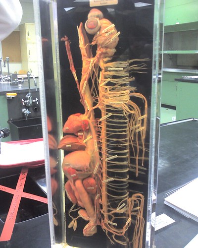
We did the entire spinal cord in one night in neuroanatomy lab. Granted, from a gross dissection point of view, a lone spinal cord is not much to look at. But when you consider the rather extensive innervation it becomes quite a daunting task.

Which organs, muscles, and body parts are innervated by which nerve tracts? How many interneurons for this system and what kinds of reflexes are involved? What will sympathetic or parasympathetic activation lead to? Which lamina of the vertebrae are receiving this type of input?

For those of you with some bio/neuro, recall that activation must also involve some reciprocal inhibition, so even a basic (monosynaptic) stretch reflex stimulates a cascade of firing.

I think next week we move up the brainstem to the medulla (mylencephalon). And we'll get our exams back. Ick.

24 September 2007

Neuroanatomy


The first neuroanatomy exam is Wednesday so I am, of course, furiously trying to cram names like sulcus limitans and stria habenularis into my brain. I labeled photographs, I have made lists of key terms, I have flipped through slide sets and I have read the textbook and somehow this subject flummoxes me. Well, not the material itself, but rather, what is the best and most efficient way to study it?

It is great practice for medical school, where will have to learn more than just neuroanatomy. If I can figure out a strategy for this class then maybe I'll be a step ahead in general anatomy. If.

07 September 2007

I deserve a cookie.


A moment of victorious immodesty: I just cranked out a 115 page IRB protocol for constraint-induced movement therapy in 4.5 days.


update: It was pushed to the October 3rd review. : (

30 July 2007

Playing neurologist

I shadowed Dr. L in clinic recently and it turned out to be particularly eventful. The medicine itself is always interesting, but this time it was the patient's behavior that made it memorable. We saw a girl who was recovering from stroke for a routine follow-up examination. Throughout the exam she was staring straight at me and refusing to acknowledge a single question or command posed by Dr. L. After trying one last time to get her to follow his finger with her eyes, he sat down and looked at her. She finally looked him in the face, pointed an arm straight out at me and said "I want her to do it." Dan looked at me, nodded, and I walked over and stood in front of the girl. I have seen at least fifty basic neurologic exams performed and could describe it in lurid detail, but standing there performing it was absolutely nerve wracking. Dr. L was standing immediately behind me interpreting everything I was doing... I was in no way evaluating the patient; I was simply the body she interacted with.

The next patient was another female, clearly somewhat on edge. Dr. L introduced me and a visiting physician (also shadowing) and began a conversation with her. Three sentences in she announced "I know you don't think I'm going to discuss my business with all these people in the room." I promptly offered to leave to make her more comfortable, but she countered, "you can stay, but the other one has to go." The visiting physician left the exam room (I would later learn she was uncomfortable with men, as the visiting physician was male, it was simply a matter of gender). She began telling a (rather sad) story about recent events and while she was talking she curled up into a ball on the exam table. When Dr. L turned to get a pen from the desk, she hopped off the table, ran across the room to me, and gave me a bear hug. After a few minutes she released me and sat in my lap. The rest of the visit was conducted with her on my lap, one arm around my shoulders.

Most of Dr. L's patients have met me once, many of them at least twice now. Quite a few remember me when they come in and ask me how school is going, which is really quite remarkable to me. After all, I am silent most of the time, just watching and smiling. Apparently though, they are beginning to feel comfortable with me, which is a great feeling. I hope this carries over to my future career, that my patients feel they can trust me and be open the way these two girls were. The way all Dr. L's patients are with him.

14 June 2007

My very own brain

This, to the left, is my brain. As in, the one in my head - the one I use every second of every day. Cool.

For those of you with no neuro background, let's name some structures.

1 Cerebellum
2 Medulla oblongata
3 Pons
4 Pituitary (right above the point of the 4)
5 Cortex
6 Splenium
7 Fornix
8 Septum pellucidum
9 Corpus callosum
10 Meninges: dura, arachnoid, pia

For the record, I have a normal brain for someone my age. The funny dark patch on the top of my head is cerebral spinal fluid (CSF), which is visible because the interhemispheric fissure was not fully perpendicular.

18 March 2007

Monthly clinic

On Friday I shadowed the outpatient neurology clinic, as I do once a month, and observed a days worth of follow-up visits. Most clinic days seem to show a theme - more likely because something registers in my subconscious than any trick of patient scheduling. Considering my recent review of genetics, it's perhaps not overly mysterious that this month impressed upon me the phenotypic markers that can signal an underlying neurologic process.

Most of you are probably familiar with some of the more famous neurologic phenotypes. For example, the Down's syndrome features of a single transverse palmar crease (simian crease), epicanthic eyelid fold, flattened nasal bridge, shortened limbs, proruding tongue, and white spots on the iris (Brushfield spots). The genetic correlate of Down's is whole or partial trisomy 21.

There are, in fact, a large number of genetic or neurologic processes that can be identified by phenotypic markers. An non-genetic example would be a perinatal (near time of birth) stroke patient. His stroke included part of the internal capsule and thus affected the neuronal tract that includes motor neurons. His stroke was right-sided, so one would expect left-sided motor affects. Indeed, because the stroke was not immediately noticed during his infancy (most infants do not get MRIs) his left side failed to develop properly. His left arm is shorter than his right (atrophy) and displays abnormal tone and fine motor control. In this case, the morphologic presentation of a shortened arm with abnormal tone allows a neurologist to begin localising his stoke before ever seeing an image.

A note about fine motor control - I get a lot of questions about this. Gross motor contol is large movements: walking, throwing, and lifting. Fine motor is things like tieing shoes, buttoning buttons, and writing. One test you might see a neurologist perform is asking a patient to touch their first finger to their thumb, then their second, third, and fourth. Another is to tap their first finger and thumb together as rapidly as possible.

Regarding motor development in infants... like anything else it's hardest to assess in younger children. Babies have little to no purposeful motor control so assessment usually consists of examining involuntery movement for abnormal posturing, tone or lack of movement. Mild deficits can be difficult to note until a child fails to develop on a normal timeline (ie, cannot stand independently or sit-up on time).

And now a genetic example. I should mention that this patient has not yet been conclusively diagnosed, we simply added Wolf-Hirschhorn to the differenital because of the phenotype. There were two children who came into clinic after having been neglected by their biological parents. It was impossible to tell whether they were delayed due to lack of input or whether there was genuine neurologic impairment of some kind. The children were placed in foster care and came back speaking 3 word sentences (they are 3 and 4 yrs old), but still underweight and incapable of tasks such as putting on clothing or drinking from normal cups. The younger child displayed tremors (attributed to more than being nervous because it affected her head as well) and the older child has "greek-helmet head", microcephaly (small head), and shortened stature. Greek helmet head is characterised by a high hairline and a broad, flat nose.

If the older child does have Wolf-Hirschhorn (deletion on chromosome 4) he's both lucky and unlucky. Unlucky because it's associated with fairly profound mental retardation, and lucky because his phenotype is (apparently) relatively mild. More extreme symptoms can include cardiac septal defects, poor development of secondary sex characteristics (genitals), renal (kidney) malformation, malrotation of the intenstines, and hand/foot contractures.

One of the important notes about these phenotypic varients is that in order to dianose anything they generally occur in clusters. For example, just having wide-set eyes or a high hairline doesn't mean you have a genetic defect or a neurologic symdrome. Cleft palate is a midline closure defect - a characteristic of Wolf-Hirschhorn syndrome. However, far more babies are born with cleft palate than Wolf-Hirschhorn: about 1 in 600-800 vs. 1 in 50,000.